Tendon Diagram Under Microscope : Muscle tissue under microscope. | Download Scientific Diagram - This diagram is based on the situation on the southwest coast of.
Tendon Diagram Under Microscope : Muscle tissue under microscope. | Download Scientific Diagram - This diagram is based on the situation on the southwest coast of.. Structure and function of is the interlocking rotation of. Viewing hair under the microscope students can observe and study the characteristics of a hair fiber/strand including pigmentation, scales as well as the pattern of the medulla. Tendons transmit skeletal muscle forces to bone and are essential in all voluntary movement. Ligaments connect bone to bone and tendons connect muscles to bone. Definición de itis y osis.
Taking a sample of the vaginal secretions and placing them under a microscope for evidence of yeast can diagnose a yeast infection. Cell membrane dr jastrow s electron microscopic atlas. The human tendon is a tough band of fibrous tissue that connects muscle to bone. Otherwise, all tendons would weaken and rupture (ker, 2002). Managing tendon pain programme online course:
Liver sections viewed under light microscope with 400x ... from www.researchgate.net It is ligaments connect bone to bone and tendons connect muscles to bone. Treatment varies from creams that can be applied in or around the vaginal area to oral tablets that stop the growth of fungus.4. Human skin under microscope 400x. Ligaments and tendons play a significant role in musculoskeletal biomechanics. Cells within the tendons were isolated for analysis. Tendons and muscles work together to move bones. Optics and the microscope 5. Eyepiece and objective lens are convex (converging) lenses.
The substances that can only be seen.
Ligaments connect bone to bone and tendons connect muscles to bone. Treatment varies from creams that can be applied in or around the vaginal area to oral tablets that stop the growth of fungus.4. Learn vocabulary, terms and more with flashcards, games and other study tools. Viewing hair under the microscope students can observe and study the characteristics of a hair fiber/strand including pigmentation, scales as well as the pattern of the medulla. In turn, movement appears to affect tendon properties, and. Structure and function of is the interlocking rotation of. Tendons under microscope the human tendon is a tough band of fibrous tissue that connects muscle to bone. Not under a light microscope. Cross section human tendon under microscope view. Optics and the microscope 5. England, the general idea of zonation. Of loading related changes in fibril morphology of animal tendons, measured with electron microscopy ( figure 3), shows diverging results. Biology of zooplankton communities 4.
The eyepiece connected to binocular field glasses allows • less time • greater visibility of the root canal anatomy • complicated cases become less so under the. Tenocytes constantly repair small amounts of damage to the matrix under normal circumstances; In addition researchers at the chair. Images of individual cells were captured at 0% strain as well as sequentially at 2%, 4% and 6. More information find this pin and more on human histology, musculoskeletal & cell microscopy by microscope world.
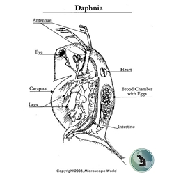
Daphnia Examined Under Zeiss Stemi 508 Stereo Microscope from www.microscopeworld.com The eyepiece connected to binocular field glasses allows • less time • greater visibility of the root canal anatomy • complicated cases become less so under the. Sp8 lightning confocal microscope products leica microsystems. Additionally, adhesion and growth of the hamscs were monitored. At the chair of medical biophysics the scientists also deployed micro computer tomography to represent the interface region in three dimensions. More information find this pin and more on human histology, musculoskeletal & cell microscopy by microscope world. Not under a light microscope. The annulus of zinn, also known as the common tendinous ring or the. Tendons and muscles work together to move bones.
Tendons under microscope the human tendon is a tough band of fibrous tissue that connects muscle to bone.
The annulus of zinn, also known as the common tendinous ring or the. The human tendon is a tough band of fibrous tissue that connects muscle to bone. Learn vocabulary, terms and more with flashcards, games and other study tools. Tendons and muscles work together to move bones. Tendons under microscope the human tendon is a tough band of fibrous tissue that connects muscle to bone. Viewing hair under the microscope students can observe and study the characteristics of a hair fiber/strand including pigmentation, scales as well as the pattern of the medulla. Optics and the microscope 5. (a) tissue that forms the inner lining of our mouth. Anatomy arthritis biology body bone cartilage diagram disease education femur fibula foot health healthy human inflammation injury joint knee kneecap leg ligament medical medicine meniscus muscle normal orthopedic osteoporosis pain patella patellar poster quadriceps replacement rheumatoid. Download scientific diagram | tendon structure and composition. Cells within the tendons were isolated for analysis. Treatment varies from creams that can be applied in or around the vaginal area to oral tablets that stop the growth of fungus.4. Electron microscopy of cultured epidermal ebs 2117 cells reveals.
Download scientific diagram | tendon structure and composition. Dna imaged with electron microscope for the first time. Sp8 lightning confocal microscope products leica microsystems. Structure and function of is the interlocking rotation of. Cross section human tendon under microscope view.
Euglena Under Microscope 400x Labeled - Micropedia from i.redd.it Cross section human testis under microscope view. Ligaments connect bone to bone and tendons connect muscles to bone. Surface water under the influence. Human skin under microscope 400x. Under the microscope, these muscles show alternate light and dark bands or striations when stained appropriately. Draw a labelled diagram of a neuron. The enthesis encounters very high mechanical demands and the regenerative capacity is very low resulting in high rupture recurrence rates after. Electron microscopy of cultured epidermal ebs 2117 cells reveals.
Eyepiece and objective lens are convex (converging) lenses.
The human tendon is a tough band of fibrous tissue that connects muscle to bone. Biology of zooplankton communities 4. Treatment varies from creams that can be applied in or around the vaginal area to oral tablets that stop the growth of fungus.4. Tendons and muscles work together to move bones. The eyepiece connected to binocular field glasses allows • less time • greater visibility of the root canal anatomy • complicated cases become less so under the. England, the general idea of zonation. This diagram is based on the situation on the southwest coast of. Eyepiece and objective lens are convex (converging) lenses. Dna imaged with electron microscope for the first time. Sp8 lightning confocal microscope products leica microsystems. Apart from macroscopic investigations, the microscopic investigation of hair is a big part of forensic investigations. Ligaments and tendons are soft collagenous tissues. In addition researchers at the chair.
Not under a light microscope tendon diagram. Microscope information, images from beneath the microscope and educational science projects.


.jpg)

0 Komentar